by
Gus Iversen, Editor in Chief | December 26, 2023
3: Can you tell me about the learning curve for sonographers trying to incorporate opto-acoustic imaging into their exams?
For the sonographer, the learning curve is much like any new device that is brought in that has a different interface and knobology than what they are used to using. Getting used to the new interface is likely the longest learning curve, but most adapt easily after a few patients. Because this is a new modality for sonographers, we want to train them equally as much in understanding this new modality and what they are looking at to better provide the radiologist with the best video clips of the patient’s anatomy and functional information from the opto-acoustics [laser] component of our probe.
The training was created by Dr. Tom Stavros the world’s leading breast ultrasound radiologist, who wrote the textbook on breast ultrasound for sonographers and radiologists. The sonographer is provided specific modules to review to educate about the new modality in advance and then on-site training by Seno’s Clinical Application’s specialist that reviews laser safety, device, and scanning, and observes and supports the sonographers with several patients.
4: Given the advantages of OA/US for reducing unnecessary biopsies and bringing value to imaging, is it reimbursed differently than conventional breast US?
Today every OA/US exam will bill their typical diagnostic ultrasound codes and if they see a suspicious area that is likely BIRADs 3 or above, turn on the laser and bill for that portion of the exam. Since, we are early in our commercialization of this new technology we have an APC code and a CPT III code that will go into effect in January 2024 that is specific to the OA exam.
Seno also has employed a third party, Pinnacle Health Group, to help our customers and patients navigate the reimbursement process including a customer hotline for support, coding materials, medical letters of necessity, etc.
5: Do you anticipate other applications for combining ultrasound with opto-acoustic imaging?
Today our technology is only approved for diagnostic breast imaging. However, previous proof of concepts and successful feasibility studies are complete in thyroid cancer and in the area of monitoring neoadjuvant care for breast cancer. The plan is that this new modality will be a platform technology for cancer diagnostics and impact other areas that today suffer from large false positive rate of biopsies.
For example, another area, like breast cancer and thyroid cancer, that tends to generate significant negative biopsies is the area of prostate cancer. Seno has developed a prototype probe for this application and has done preliminary work in investigating what is needed for a feasibility protocol.
In some of the aforementioned disease states, Seno anticipates that our study work will likely be in the area of determining aggressive cancers versus those that are more watchful waiting; or whether in the area of neoadjuvant care we can determine after the first chemo regiment if the treatment is working or not. From a patient and healthcare cost perspective Seno believes this will be our greater impact. Each of these applications have significant unmet needs and large market opportunities.
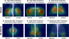
At the San Antonio Breast Cancer Symposium this past week, the results from our neoadjuvant chemotherapy feasibility study at University Texas Southwest with Dr. Basak Dogan, was presented. This presentation was well attended, the Imagio OA/US system shows promise in correlating pathological response to neoadjuvant therapy even as early as the first regimen. One can imagine how important this would be for patients to know as early as possible, so they can change their treatment options much earlier, saving patients time, physical and mental stress, and cost.
The Imagio Breast Imaging System was approved by the FDA in January 2021 with additional approval in June 2022.
The study was published in
Academic Radiology.
Back to HCB News