New algorithm shows if — and why — cancer patients should undergo biopsies
by
John R. Fischer, Senior Reporter | January 26, 2022

A new AI algorithm has been trained like a radiologist to show how it diagnoses malignant and benign breast tumors
A new algorithm developed at Duke University is designed to not only help indicate if a cancer patient should undergo an invasive biopsy but shows radiologists how it reached its conclusions.
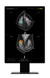
Designed to locate and evaluate potentially cancerous lesions in mammograms, the AI solution is trained just like a radiologist would be to assess tumors, and can show the evidence it used to make its findings. This involves assessing the edges of lesions, rather than surrounding tissues, or learn based on how the specific equipment that it is used with functions. As a result, it can freely develop its own procedures.
Additionally, it was trained with a validation data set of 1,136 images. The platform classifies a breast lesion into a specific category. Radiologists can then assess if the AI has based its decision on viable criteria. If so, the classification can be confidently included in the radiologist’s report. If not, the radiologist can override the AI and understand why it failed.
Most independent algorithms for medical imaging are trained on less than 1000 scans or contain demographic information. This, along with recent failures around the use of these solutions, has led many physicians to question their use in high-stakes medical decisions.
“By suggesting an interpretable diagnosis, the algorithm can help radiologists to improve their performance and consistency. Long term, we hope that this approach can avoid many benign biopsies, which would benefit not only the patients but also expedite the time-consuming processes of diagnostic workup and biopsy scheduling,” Joseph Lo, professor of radiology at Duke University School of Medicine, told HCB News.
The aim, according to Lo and his colleagues, is for their approach to reduce the number of benign biopsies. This would not only benefit patients but expedite the time-consuming processes of diagnostic workup and biopsy scheduling.
Images were taken from 484 patients at Duke University Health System. Using them, the researchers taught the algorithm to find the suspicious lesions in question and ignore the health tissue surrounding it and other irrelevant data.
Images were labeled to teach it to focus on the edges of lesions, where the potential tumors met healthy surrounding tissue, and compare them to edges in images with known cancerous and benign outcomes.
Radiologists often look for radiating lines or fuzzy edges known as mass margins first when assessing tumors, as they are the best predictors of cancerous breast lesions. Also, cancerous cells replicate and expand fast, which makes it hard to see all of a developing tumor’s edges in mammograms.
|
|
|
You Must Be Logged In To Post A Comment
|